Detraining, Confusion and the Blood-Brain Barrier
What happens when we stop exercising? How the WHO discredited themselves? And how to reconstruct the blood-brain barrier?

This week I’m trying out a slightly different approach. I’ll try to keep myself bound to 3 topics and try to explore different perspectives around them. By all means, let me know what works for you.
Detraining
The main benefits that aerobic exercise has on our bodies are fat loss, improving our cardiovascular system, improving VO2max. I think we all agree that these are all “indicators” of a healthy body. Now let’s look at what happens when we stop exercising, also known as detraining.
VO2max (the rate at which oxygen is consumed in tissues) starts significantly decreasing after 2-4 weeks (6%), which is related to a decreased cardiac output and decreased blood volume. After 9 weeks it decreases by 19% and by 11 weeks by 20-25%. Luckily, all these losses can be made up with a few weeks of good aerobic exercise.
From a cardiovascular standpoint, the heart starts losing its muscle, which decreases its contractility. This results in higher heart rate and recovery heart rate.
Some interesting things also start happening on the metabolic level. The respiratory exchange ratio goes up, which is an indicator of inefficient fat oxidation. This, in turn, leads to fat accumulation. Of course, lactate builds up faster and less glycogen is stored in the muscles. Unsurprisingly, the muscle mass and the number of type 1 muscle fibres also decreases.
There’s a lot more to explore here and this article does just that. In general, the downside of not being physically active in any way is substantial. What is the upside? More time? More rest? More energy? In my experience, it’s quite the contrary. Better sleep, efficiency and overall well-being. The upside is huge.
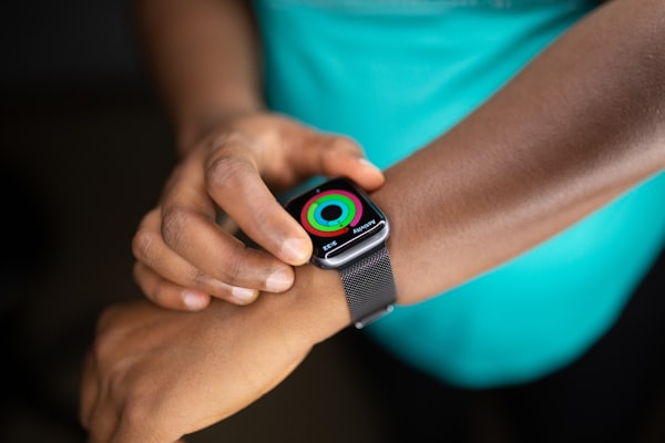
Personal example: My VO2max correlates almost perfectly with medical school. According to my Garmin Vivoactive 3 it started at about 55 ml/kg/min in June 2018 and it fell to 42 ml/kg/min until January 2019 - that’s when I started medical school. Having realised that a 25% drop is quite significant, I started working out and running and by June 2019 I was at 48 ml/kg/min. The same happened this year. But I also have some more recent data, that shows the effect of exercise (resistance and aerobic training). I was at 43 ml/kg/min in January and am now at 47 ml/kg/min. I’m sure that if I maintained a more regular aerobic exercise, this number would be higher, but it's awesome to see some feedback.
Speaking of aerobic exercise I noticed something remarkable when running. In the past, my goal would be to have the highest pace possible…usually a little less than 5 min/km. That was okay, but I couldn’t endure long distances, nor did I enjoy running. But then I reconsidered, lowered my pace and kept my heart rate between Zone 2 and Zone 3 - so about 70% of my maximal heart rate. All of a sudden running became a pleasant experience that I’m now using to get some fresh air and also improve my VO2max. Kind of like Derek Sivers describes in this post.
Faster, more and more often is not necessarily better. Especially if you’re not enjoying it.
Confusion
The World Health Organization (WHO) said on a press conference that COVID-19 is “very rarely transmissible among asymptomatic patients”. They clarified the statement later, but how did we get here?
Throughout the pandemic and before it started, WHO was giving out some misleading statements and actions. At first, there was no evident human-to-human transmission. Then, after officials in Wuhan quarantined 57 million people, they needed another week to call it a global health emergency. They were getting the information from China, who were misinforming the world about the virus. This is now clear. And yet, WHO was praising its actions just before they finally called it a pandemic.
The WHO, an organisation that the entire world relies on, makes confusing statements and fails to respond on time. If they were so careful not to call it a pandemic, why weren’t they more careful when they reported there’s no evidence of human-to-human transmission?
Fast forward to June 8 and the WHO press conference. Among other things, they stressed the fact that the most important thing is that each individual knows what to do to protect themselves and those around them. Plus, they warned that this pandemic is not over. The statement that sparked confusion was that the asymptomatic transmission of COVID-19 was “very rare”. The following day they walked back on that statement and said this isn’t their policy, but that these were results of some not yet peer-reviewed studies.
Of course asymptomatic and presymptomatic have different meanings. We have enough data to know that presymptomatic people infect others (paper from Nature). And we know that about 40-45% of all infections are asymptomatic (paper from Annals of Internal Medicine), but there isn’t enough data yet to support their claim.
This may be “very rare” in the end, but why make such a claim? The studies are not yet peer-reviewed and there’s a significant body of knowledge that indicates otherwise?
There’s no question there are top-notch scientists and doctors at WHO, but the way they communicated during the pandemic was not exemplar. For one, they’re responsible for almost 8 billion people and their statements mean too much to make them confusing or even wrong. Why would you want to give out confusing statements? As if the protests all around the world aren’t enough for the virus to continue to spread and (most likely) cause a second wave. As if the horrifying situation in South America (and now Africa) isn’t enough. And there’s no vaccine yet.
Speaking of vaccines, let’s shift to more a pleasant topic: Horseshoe Crabs (they look like this). I never imagined such a connection. Horseshoe Crabs contain a component called LAL that’s used in drug development to test for bacterial toxins - endotoxins. “Pharmaceutical companies must make sure the toxins are not present in any injectable drugs they make. Ingredients, like water, must be tested at each step of the manufacturing process, as well as in the final product.” There are of course scientific considerations on how to replace the need to use LAL from a live animal. A synthetic equivalent, recombinant factor C (rFC) already exists and is already used in Europe. But not yet in the USA.
Blood-Brain Barrier
Researchers reconstructed the human blood-brain barrier (BBB) Medicine and looked at the effect amyloid deposits (common in Alzheimer’s disease) have on its function.
In a few issues to date, I discussed findings in various papers that are connected to Alzheimer’s disease. Some of them by testing patients’ blood instead of CSF and others that did metabolomic studies of the CSF.
The blood-brain barrier (BBB) is a structure along the brain’s vasculature that shields the brain against pathogens. It’s critical for proper neuronal function and tightly regulates what goes into the brain’s circulation. 3 structures build it: pericytes, brain endothelial cells and astrocytes.
Alzheimer’s disease (AD) is known to cause accumulation of protein plaques in the brain’s circulation. These proteins also affect the function of the BBB and make it less efficient. This is known as cerebral amyloid angiopathy (CAA) and it accelerates cognitive degeneration.
There is another predictor when it comes to AD and CAA, whose molecular mechanism is unknown. This is the notorious APOE4 gene, which strongly correlates with the development of AD. Because we don’t know the molecular mechanisms with which it causes CAA and AD, we can’t treat it or properly change our lifestyle to prevent it.
In the mentioned paper they interestingly propose a missing piece of the puzzle. This is a component called calcineurin. It’s a calcium and calmodulin-dependent serine/threonine protein phosphatase and activates the T cells of the immune system. It turns out that dysregulation of calcineurin induces APOE4-associated CAA. On the other hand, if calcineurin is inhibited, the CAA is reduced and fewer plaques are formed in the brain vasculature. This provides new insight into the underlying mechanism and once again offer a target for drug development.


